Optical tracking of membrane potential in cell cultures
Genetically Encoded Voltage Indicator
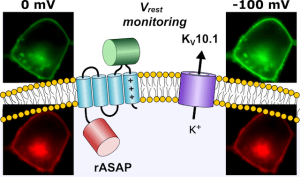
Illustration: FSU BiophysikThe electrical membrane voltage (Vm) is a decisive characteristic of excitable and nonexcitable cells as it determines direction and magnitude of ion transport across the membrane. Galvanic measurement of Vm, however, requires the access of an electrically conducting microelectrode to the cellular cytosol. Although routinely applied in various electrophysiological methods, this approach is not only severely invasive but also tedious and time-consuming, thus precluding the automatic evaluation of many cells in parallel. Here we present optimized genetically encoded voltage indicators that operate in a ratiometric fashion. Applied to cell cultures, compound Vm of several thousands of cells is readily measured. Furthermore, we show how ectopic expression of K+ channel mutants that cause neurodevelopmental disease such as the Temple-Baraitser syndrome (KV10.1) hyperpolarizes the cells. The introduced method also allowed monitoring the speed with which triggered expression of genes coding for K+ channels lead to an alteration of Vm.
